One of our requirements for the first semester is to
complete a two-day rotation in the histology lab. The rotation entails waking
up before the sun rises and observing the histotechs embed and cut
specimens. The histotechs have to start
work super early in order for everything to be ready by the time the
Pathologists arrive. When my alarm went
off at 3:45am I couldn’t help but wonder how anyone could be fully functional
at that hour. Luckily we do the
rotations with another classmate, which made being up so early way less
painful (thanks Corey!).
 |
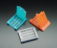
| Cassettes |
Histology means the study of tissues. The histotechnologists’ job
begins after the PAs take sections of a specimen and submit those sections in cassettes. The cassettes are delivered
to the histology lab and each one is scanned into the computer, in order to
keep a log of all the cassettes that are received. The histotech then takes the tissue and embeds
it in paraffin; they must have
superhuman fingers because the wax is hot!
 Once the paraffin solidifies, they
cut thin sections of the tissue on a microtome. They place the ribbon
of sections in a water bath and then pick up the sections on a microscope
slide. They create as many slides as
needed based on the different stains that are required for the specimen. After the tissue is on the slides, the slides are
stained and coverslipped (at our lab they have an automatic staining and
coverslip machine). Once that is finished, the histotechs gather the report and the slides and give them to the
pathologists to review under the microscope.
 |
| Paraffin block being cut on microtome |
After watching the whole process, Corey and I were able to
try embedding and cutting various sample specimens. When we came back for our
second day of rotations the histotechs had stained our slides and we were able
to view them under the microscope.
 |
| Some of the slides that I made |
As we are not far in our microanatomy class, we had fun
trying to describe what we saw under the microscope as if we were the residents
during a conference. “On high power we see a lot of fibrous tissue and enlarged
nuclei.” It’s funny how the slides just look like pretty pink and blue blobs to
me right now, but with time I’ll be able to recognize certain diseases based on
these microscopic examinations.
My favorite thing that I’ve seen under the microscope so far
is an eccrine duct (it looks like a 5 year olds art work!):

This was really interesting and helpful. Thanks for including photos :)
ReplyDelete