Sunday, February 24, 2013

Histology Rotation


One of our requirements for the first semester is to complete a two-day rotation in the histology lab. The rotation entails waking up before the sun rises and observing the histotechs embed and cut specimens.  The histotechs have to start work super early in order for everything to be ready by the time the Pathologists arrive.  When my alarm went off at 3:45am I couldn’t help but wonder how anyone could be fully functional at that hour.  Luckily we do the rotations with another classmate, which made being up so early way less painful (thanks Corey!).
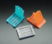
Cassettes
Histology means the study of tissues. The histotechnologists’ job begins after the PAs take sections of a specimen and submit those sections in cassettes. The cassettes are delivered to the histology lab and each one is scanned into the computer, in order to keep a log of all the cassettes that are received.  The histotech then takes the tissue and embeds it in paraffin; they must have superhuman fingers because the wax is hot! 

Once the paraffin solidifies, they cut thin sections of the tissue on a microtome. They place the ribbon of sections in a water bath and then pick up the sections on a microscope slide.  They create as many slides as needed based on the different stains that are required for the specimen.  After the tissue is on the slides, the slides are stained and coverslipped (at our lab they have an automatic staining and coverslip machine).  Once that is finished, the histotechs gather the report and the slides and give them to the pathologists to review under the microscope.

Paraffin block being cut on microtome
After watching the whole process, Corey and I were able to try embedding and cutting various sample specimens. When we came back for our second day of rotations the histotechs had stained our slides and we were able to view them under the microscope.
Some of the slides that I made

As we are not far in our microanatomy class, we had fun trying to describe what we saw under the microscope as if we were the residents during a conference. “On high power we see a lot of fibrous tissue and enlarged nuclei.” It’s funny how the slides just look like pretty pink and blue blobs to me right now, but with time I’ll be able to recognize certain diseases based on these microscopic examinations. 


My favorite thing that I’ve seen under the microscope so far is an eccrine duct (it looks like a 5 year olds art work!):

Saturday, February 16, 2013

Frozen Sections


Last Sunday we had our first frozen section lab.  Frozen sections are a big part of the PA’s duties.  I've heard people say it is one of their favorite parts about being a PA, but right now it seems a little daunting.   The frozen section is an intraoperative procedure. Since the patient is still on the table in the OR, the pathologists’ assistant has to work fast.  The CAP regulations say that frozen section procedures must be done in 20 minutes.  This includes receipt, accessioning, examination, any inking or special procedures, sign out, and call in to the surgeon.  Therefore the duties that the PA performs must be completed in 7 minutes.  

Here is a description of the PA’s duties (I’ll try to make it as least technical as possible):

Cryostat
Once we receive the specimen we select the tissue that we will use for the frozen section (no necrotic tissue, no normal tissue, we want to demonstrate as much information as possible in 1-2 slides). We ink the margins if necessary and then begin our frozen section procedure.  The frozen section procedure takes place in the cryostat, a chamber that is at least -20°C.  

The tissue is placed on a chuck and surrounded by OCT (essentially a goo that freezes). 


Chucks
The chuck is put into the head of the microtome  and then we turn the wheel on the side of the cryostat to start taking our sections.


Chuck and selected tissue on microtome

Turning the wheel moves the microtome closer to the knife blade. Each advancement of the wheel cuts a 5 micron section of tissue (this is adjustable, but for our purposes we will keep it at 5 microns). Once it looks like the whole face of the tissue is shown, we use a brush to pull the section away from the block to place it on a slide.


Using the brush to remove the section
The tricky part with this step is getting the section to be smooth so that the tissue doesn’t fold up on itself.  Once the section is on the slide we fix it in alcohol and stain it so that specific components can be seen easily by the pathologist when looking through the microscope.

And that is essentially the frozen section! Can you imagine doing all of that in only 7 minutes? 

         
During our lab we practiced on pieces of hotdogs.  I was very slow at first, but I started to get the hang of it after a couple of tries.  We didn’t time ourselves, but I’m sure I’m no where near the 7 minute limit.  But with some more practice and many hotdogs later I should have the hang of it!

*These pictures are not my own, they were obtained from google.


On a non related note, my grandparents sent me a card for Valentine’s day with this drawn on it: 

My grandma did a great job and I thought it was adorable!


Tuesday, February 12, 2013

Busy Week

I apologize for the lack of an update this week.  I've had a lot of reading to complete in preparation for an exam on Thursday.  Also my histology rotation is this week which requires me to be functional at 5am.  I would love to give an update, but obviously school comes first.  Look out for a post about our first frozen section lab and my histology rotation in the near future!

Monday, February 4, 2013

This Week the Trend...EXAMS!


I’ve noticed that I’m already bad about posting regularly (I promise to try to get better).  It is hard to believe that the fourth week of the PA program is starting. We are just finishing up our first round of exams: Anatomical Techniques was last Monday, Anatomy was Friday, General Pathology was today, and our Anatomy Lab Practical is Wednesday.

Here’s a rundown of what the tests are like:

Anatomical Techniques- A mix of short answer and essay type questions with a few multiple-choice questions. For this exam you really had to know the material; which makes sense because we need to know all this information for when we are practicing PAs as well. 

Anatomy- The first half of the test were multiple-choice and fill in the blank style questions based off of the lectures. The second half consisted of labeling anatomical plates. To the right is a plate for the next exam.  We are given about 10 of these plates to learn before each exam and then 3-4 are chosen for us to label during the exam.  For the last test I started learning these plates 4 days before, which wasn’t an adequate amount of time.  My strategy now is to learn a plate a day so that when exam day comes I’ll be much more confident in the plates. 
Anatomy is straightforward and is pretty much memorization based.  I’ve always been good at memorizing things quickly. When I was little I loved to play “Guess Who?” (anyone remember that game?) with my dad.  The premise of the game is to guess your opponent’s mystery person before your opponent guesses yours. By the end of the game I could recall which characters we had already used and in what order; so that I could immediately guess which character my dad had in his hand.  In a way, the anatomy exams are like a very intense game of “Guess Who?”.

Pathology- This exam was more similar to the exams in undergrad.  It was completely multiple-choice and based directly from the lecture notes.

Last week taught me a lot about time management while in the program.  Although I feel that I did decently on the tests, I know that I need to change my study strategies for the future.  Studying for tests in grad school is completely different from undergrad.  You definitely cannot wait until the day before a test to start studying; it won’t end well. Also I should mention that we do a TON of reading (articles, textbooks, procedures, etc.) You can’t get stressed about the readings or studying for exams; you just have to accept it and make it routine.


On a more exciting note, we have also been attending various pathology conferences.  So far we’ve gone to ones regarding autopsies, brain cutting, the pancreas, and gross dissection.  At the conferences the Pathologists and the residents take turns presenting a case and then state their findings.  It reminds me of Grey’s Anatomy when they do rounds (yes I just compared real life to a TV show). The conferences are completely over my head right now, but I still think it is a great experience.  I cannot wait for the day when I know just as much about pathology.